tree in bud opacities pneumonia
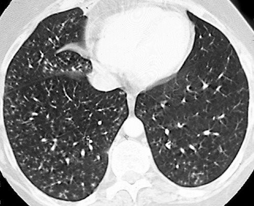
It consists of small centrilobular nodules of soft-tissue attenuation connected to multiple branching linear structures of similar caliber that originate from a single stalk. On chest CT the tree-in-bud opacities had vanished.

Tree In Bud Pattern Radiology Case Radiopaedia Org
We here describe an unusual cause of TIB during the COVID-19 pandemic.
. Thus pulmonary infiltrates can be mistaken as pneumonia as patients are considered immunocompromised. The tree-in-bud pattern suggests active and contagious disease especially when associated with adjacent cavitary disease within the lungs. Although mycoplasma pneumonia is typically characterized by patchy reticulonodular and ground-glass opacities it can also be manifested by centrilobular nodules and tree-in-bud nodularity.
Tree-in-bud TIB appearance in computed tomography CT chest is most commonly a manifestation of infection. Originally reported in cases of endobronchial spread of Mycobacterium tuberculosis this pattern is now. Three months later repeat chest radiography revealed worsening of the bilateral lower lobe nodular opacities Fig 1C 1DA CT scan of the chest was performed that showed dense branching.
These small clustered branching and nodular opacities represent terminal airway mucous impaction with adjacent peribronchiolar inflammation. Consolidation and tree-in-bud opacities bronchopneumonia pattern were usually attributed to bacterial infection and aspiration pneumonia. The tree-in-bud pattern is commonly seen at thin-section computed tomography CT of the lungs.
Ad A Vaccine Can Reduce Your Risk of Illness By As Much as 40-60. The most common CT findings are centrilobular nodules and branching linear and nodular opacities. A young male patient who had a history of fever cough and respiratory distress presented in the emergency department.
Because the majority of candida growth in bronchoalveolar-lavage fluid is correctly attributed to colonization it. Candida pneumonia is rarely diagnosed. Tree-in-bud pattern is seen when peripheral airways are filled with pus or fluid with peribronchial inflammation.
The tree-in-bud pattern suggests active and contagious disease especially when associated with adjacent cavitary disease within the lungs. The tree-in-bud sign has been described in cases of acute aspiration 13. He had brief improvement after two courses of levofloxacin but symptoms recurred.
This may prompt inappropriate treatment. A Quick Prick Can Spare You From A Deadly Combination of The Flu Pneumonia Inflammation. BAL results were positive for Haemophilus haemolyticus and negative for malignant cells.
It is often associated with peribronchiolar inflammation29 cicatricial scarring of many bronchioles results in the indirect sign of patchy density differences of the lung parenchyma reflecting areas of. Contrast-enhanced CT computed tomography thorax revealed tree-in-bud TIB opacities. The most common CT findings are centrilobular nodules and branching linear and nodular opacities.
Four months later the patient was still in very good health and not suffering from respiratory complaints. The term centrilobular branching opacity is desirable in case the bud is absent. 87 rows the tree-in-bud sign reflects the presence of dilated centrilobular bronchioles with lumina that are impacted with mucus fluid or pus.
This tree-in-bud pattern is due to the presence of caseation necrosis and granuloma-. However after listening to patients voice and reviewing the images on CT thorax the diagnosis was confirmed as aspiration bronchiolitis. CONCLUSION The tree-in-bud pattern or sign should be used in case of visible tree and bud.
While the tree-in-bud appearance usually represents endobronchial spread of infection given the closeness of small pulmonary arteries and small airways sharing branching morphology-bronchovascular bundle a rarer cause of the tree-in-bud sign is infiltration of the small pulmonary arteries or axial interstitium 367. The tree-in-bud sign is a nonspecific imaging finding that implies impaction within bronchioles the smallest airway passages in the lung. Provisional diagnosis of pulmonary tuberculosis was made and was referred to the respiratory team.
The differential for this finding includes malignant and inflammatory etiologies either infectious or sterile.
View Of Tree In Bud The Southwest Respiratory And Critical Care Chronicles
View Of Tree In Bud The Southwest Respiratory And Critical Care Chronicles
2
2

Pdf Tree In Bud Semantic Scholar

Tree In Bud Pattern Pulmonary Tb Eurorad
2

Tree In Bud Pattern Pulmonary Tb Eurorad

Tree In Bud Pattern Radiology Case Radiopaedia Org

Pdf Causes And Imaging Patterns Of Tree In Bud Opacities Semantic Scholar

Tree In Bud Caused By Haemophilus Influenzae Radiology Case Radiopaedia Org

Tree In Bud Pattern Pulmonary Tb Eurorad
2

Co Rads 2 With Tree In Bud Sign A 27 Year Old Male Attended The Download Scientific Diagram
2

Tree In Bud Caused By Haemophilus Influenzae Radiology Case Radiopaedia Org

Hrct Scan Of The Chest Showing Diffuse Micronodules And Tree In Bud Download Scientific Diagram
Tree In Bud Sign Lungs

Chest Ct With Multifocal Tree In Bud Opacities Diffuse Bronchiectasis Download Scientific Diagram